
Ultrasound technology has become an integral part of modern healthcare. It’s revolutionizing how healthcare professionals diagnose and monitor a wide range of medical conditions.
In fact, between 2004 and 2018, 49% of general physicians utilized ultrasound.
You probably already know that ultrasound is commonly used during pregnancy for fetus growth and development monitoring. But does it have other applications? Yes, and that’s what we’re going to explore in this blog.
So, without further ado, let’s take a closer look at the several common ultrasound uses to better appreciate the technology’s immense impact on modern medicine.
How Ultrasound Works
Before we begin our exploration of common ultrasound uses, let us first gain a clear understanding of this innovative technology. So, how does it really work?
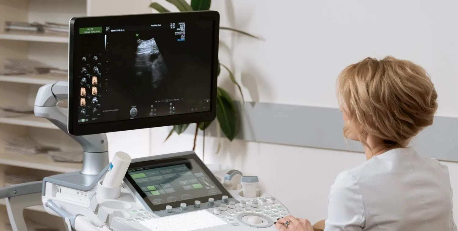
Ultrasound technology operates on the principle of sound waves. A transducer, which acts as both a transmitter and receiver of sound waves, is used to generate and detect these waves.
The transducer emits high-frequency sound waves that penetrate the body and interact with the underlying structures. When the sound waves encounter boundaries between different tissues or organs, such as the heart or liver, they bounce back to the transducer. The bouncing back of sound waves is recorded and converted into real-time images on a monitor.
Ultrasound Uses
Now, we’re ready to tackle the different uses of ultrasound technology. Here are some of them:
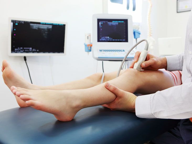
- Musculoskeletal Applications: Ultrasound enables clinicians to evaluate various joint conditions, including arthritis and ligament tears.Muscle and tendon injuries are also frequent occurrences, especially among athletes and individuals involved in physical activities. Ultrasound excels in diagnosing these injuries, too.
Another important musculoskeletal application of ultrasound is guiding injections, which is why there are now courses available for ultrasound-guided vascular access. Curious to know more about such courses? Click here or check other resources online.
- Abdominal Applications: In addition to musculoskeletal applications, Ultrasound is commonly used to assess liver and kidney function. It can evaluate the size, shape, and texture of these organs, as well as identify any abnormalities such as cysts, tumors, or fatty infiltration. The diagnosis of liver cirrhosis, kidney obstruction, and other vascular conditions also becomes possible with it.Ultrasound is also extensively used to evaluate the gallbladder and pancreas. It can identify gallstones and gallbladder inflammation (cholecystitis). Similarly, ultrasound aids in assessing the pancreas for abnormalities such as pancreatitis or pancreatic tumors.
Source: healthline.com - Cardiac Applications: Echocardiography utilizes ultrasound to assess the structure and function of the heart. It provides real-time images of the heart chambers, valves, and blood flow patterns, enabling the evaluation of cardiac wall motion, ventricular function, and chamber dimensions. Through it, clinicians can diagnose conditions such as heart valve abnormalities, congenital heart defects, and cardiomyopathies.
Of course, since ultrasound allows for dynamic visualization of the valves, it also enables the characterization of conditions like mitral valve prolapse, aortic stenosis, or mitral regurgitation, guiding treatment decisions and monitoring disease progression.
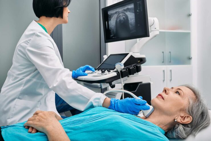
Lastly, it aids in identifying deep vein thrombosis (DVT), a potentially life-threatening condition, by visualizing blood flow in the extremities. Additionally, ultrasound can assess peripheral arteries for conditions like arterial stenosis or aneurysms. - Thyroid and Breast Imaging: Thyroid nodules are commonly detected during routine physical examinations or imaging studies. Ultrasound plays a crucial role in evaluating these nodules. It can determine their size, shape, and composition, helping clinicians categorize them as benign or potentially malignant. Not only that, but ultrasound-guided fine-needle aspiration (FNA) biopsy is also often performed to obtain tissue samples for further analysis.Ultrasound also complements mammography and clinical examination in the evaluation of breast abnormalities. It helps in distinguishing between solid masses and cystic lesions, providing additional information for diagnosis and treatment planning. Ultrasound is particularly useful in assessing dense breast tissue and young patients, as it doesn’t use ionizing radiation and can effectively visualize the breast tissue.
- Physical Therapy and Rehabilitation and Tumor Ablation: Ultrasound’s applications aren’t limited to diagnostic tests. It also has therapeutic uses. In fact, physical therapy and rehabilitation often utilize ultrasound as a valuable adjunct in the management of various musculoskeletal conditions.
The application of ultrasound waves can promote tissue relaxation, increase local blood flow, and reduce inflammation. It’s no wonder why it’s a popular choice in physical therapy settings.
In addition to its applications in physical therapy, ultrasound has a role in tumor ablation, offering a non-invasive alternative to surgery or radiation therapy. A good example is the high-intensity focused ultrasound (HIFU) that uses ultrasound waves to generate high temperatures in a targeted area, effectively destroying tumors.

Ultrasound’s ability to provide real-time imaging and its non-invasive and painless nature make it a significant component of many medical procedures. Still, its overall effectiveness depends on the skills of its operator.
The Limitations of Ultrasound
Ultrasound, despite its amazing functionalities, has limitations. For clinicians and patients alike, here are some important considerations:
- Operator-dependency: One limitation of ultrasound is its operator-dependency, which we’ve also learned in the previous section. The quality of the ultrasound images can vary depending on the skill and experience of the operator. The interpretation of ultrasound findings also relies on the expertise of the clinician. Therefore, it’s essential to have well-trained sonographers and interpreting physicians to ensure accurate and reliable results.
- Limited penetration in obese patients: Did you know that Ultrasound waves have difficulty penetrating through the increased adipose tissue in obese patients? Yes, you heard that right. And that can result in poorer image quality and reduced visualization of deeper structures. That’s why, in some cases, alternative imaging techniques, such as computed tomography (CT) or magnetic resonance imaging (MRI), may be necessary to evaluate certain conditions in obese individuals.
- Difficulty in visualizing certain structures: Some structures or conditions may be challenging to visualize using ultrasound alone. For example, air-filled structures, such as the lungs or bowel, can cause acoustic shadowing, limiting the ultrasound’s ability to visualize underlying tissues. Similarly, calcifications or dense bones can create acoustic artifacts that obscure the surrounding structures. In such cases, additional imaging modalities may be required to obtain a comprehensive evaluation.
Of course, it’s important to keep in mind these limitations when utilizing ultrasound for diagnostic or therapeutic purposes. And if needed, use other imaging modalities for the best results.
Conclusion
Ultrasound plays a crucial role in modern healthcare, providing clinicians with a safe, non-invasive, and reliable imaging modality. From its diagnostic capabilities to its applications in interventions and therapies, ultrasound continues to revolutionize the way we approach patient care. And as technology continues to advance, one can imagine how ultrasound will continue to do wonders in healthcare in the coming years!